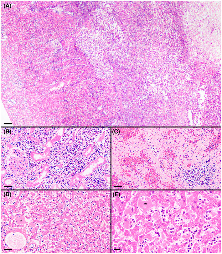
FIGURE 4.

Histologic specimens of ileum (A), renal cortex (B), spleen (C), and liver (D and E). Effacing transmural necrotizing and suppurative lesion within the ileum, with the serosa to the left and the ulcerated mucosa to the right (A). Marked suppurative inflammation within the renal cortex and associated tubular degeneration (B). Marked amyloid deposition throughout this region of the spleen (C). Moderate hepatic amyloid deposition (asterisks denote representative accumulations) and many sinusoidal leukocytes, mostly neutrophils, despite a low‐normal blood neutrophil concentration (D and E). H&E stain; magnification bars = 100 μm (A), 50 μm (B‐D), 20 μm (E)
