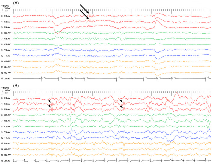
FIGURE 4.

Interictal EEGs from 2 Pomeranians that were diagnosed as IE (A, B). Both had focal seizures with limb contraction. (A) Spikes (A, arrows) and sharp waves (B, arrowheads) are seen in the frontal lobe. Note that Pz‐AV in (B) is compatible with Oz‐AV in (A). The EEG montage used for these 2 dogs was as previously described. 18 For these cases, the average reference method was used. Other recording conditions were as follows: sampling frequency = 1000 Hz; high frequency filter = 60 Hz; time constant = 0.1; sensitivity = 10 μV/mm; AC cut‐off notch filter = ON; and tracing speed = 10 s/view
