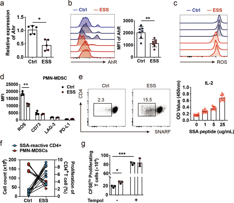
Fig. 3.
Reduced AhR expression is associated with a defective PMN-MDSC response in ESS development. a, b The expression of AhR was determined in splenic PMN-MDSCs from naïve and ESS mice by qPCR (a) and flow cytometric analysis (b). c, d Splenic PMN-MDSCs from naïve and ESS mice were sorting-purified for detection of regulatory molecules (d) by flow cytometry, and a representative histogram showed intracellular ROS production (c). e CD44+CD4+ T cells from naïve control (Ctrl) or ESS mice were sorting-purified and cocultured with SG protein-loaded dendritic cells for 24 h; rapid proliferation of SNARFlo autoreactive CD4+ T cells was detected by flow cytometry, and IL-2 production by CD44+CD4+ T cells from ESS mice was measured by ELISA in a dose-dependent manner. f Proliferative CD4+ T cells were stimulated with SG protein, and splenic PMN-MDSCs were enumerated in Ctrl and ESS mice. g Splenic PMN-MDSCs from naïve and ESS mice were sorting-purified and subjected to coculture with CD4+ T cells in the presence or absence of Tempol, and proliferative T cells were analyzed by flow cytometry. Data were derived from at least three independent experiments. Data were presented as the mean ± SD. *P < 0.05 and **P < 0.01

