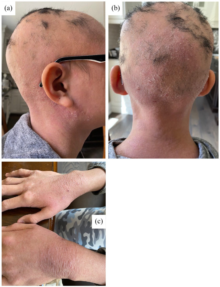
Figure 1.
Clinical photographs of the patient at first appointment. Images were captured by the patient, which were reviewed. Photos of alopecia totalis of the scalp with a few scattered tufts of hair and erythematous dry skin. (a) Lateral view shows patches of hair in the parietal vertex. (b) Posterior view shows hair patches in the occipital vertex. (c) Picture of dorsal hands with severe and active dermatitis with xerosis, scaling and erythematous lichenification bilaterally.

