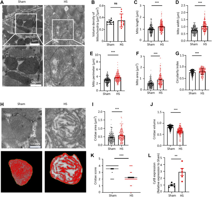
FIGURE 1.
HS damages the myocardial mitochondrial ultrastructure and increases the mtDNA release. (A) Representative transmission electron microscopic (TEM) images of mitochondria in the myocardial tissue of rats from the sham group and HS group. (B–G) Statistical results of the myocardial mitochondrial volume density, length, width, perimeter, area, and circularity index in the sham group and HS group. ImageJ was used to quantitatively analyse the myocardial mitochondrial morphology, n = 200 mitochondria from 5 rats in each group, ***p < 0.001 compared with the sham group. (H) Representative TEM images of myocardial mitochondrial cristae ultrastructure of the sham group and HS group rats. (I–K) Statistical results of the mitochondrial cristae area, cristae volume, and cristae score, n = 200 mitochondria from 5 rats per group, ***p < 0.001 compared with the sham group. (L) Statistical results of the mitochondrial DNA CytB content in peripheral blood of the sham group and HS group. n = 5 rats per group, ***p < 0.01 compared with the sham group.

