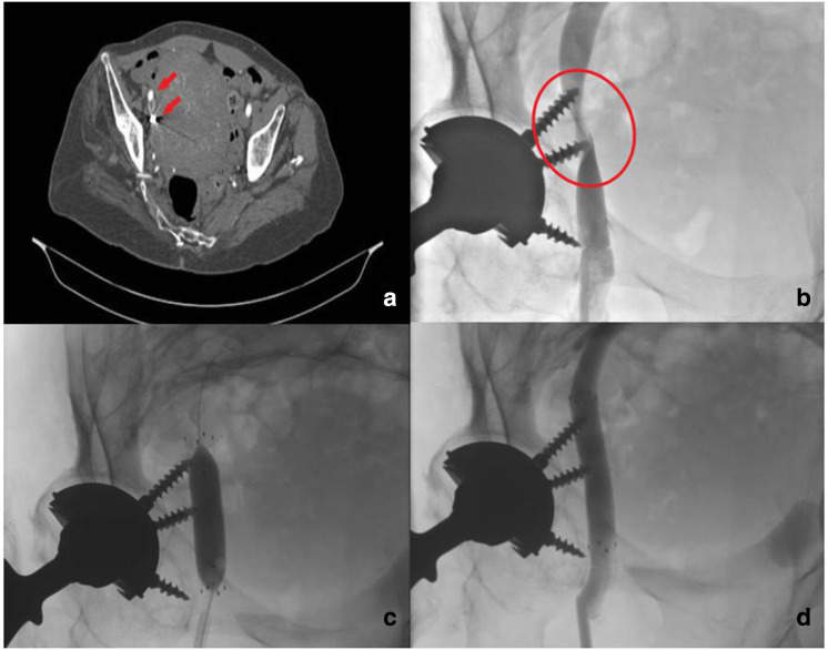
Fig. 4.
a) CT pelvis showing intrapelvic screws (red arrows) on the right side.b) Venography demonstrating iliac external vein stenosis caused by screws. c) Venography showing balloon at the stenosis site.d) Venography after stent insertion. The image shows the full flow in iliac external vein. The patient was prescribed dual antiplatelet therapy with 100 mg of aspirin and 75 mg of clopidogrel daily for four weeks since the literature suggests that this a critical period o for early stent occlusion. Before surgery, antiplatelets were suspended one week before the planned surgery.

