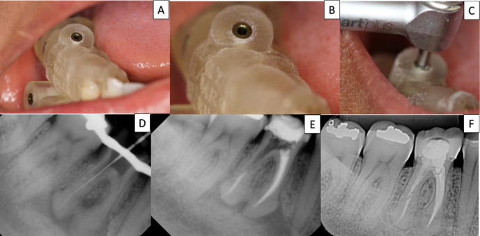
Figure 3.
A, B, C) The guide was positioned on the teeth and the adaptation and drill position verification; D) radiographic examination proves that patency patience was achieved in the distal canals; E) obturation was performed; F) digital radiography 24-month follow-up showing complete healing of the lesion

