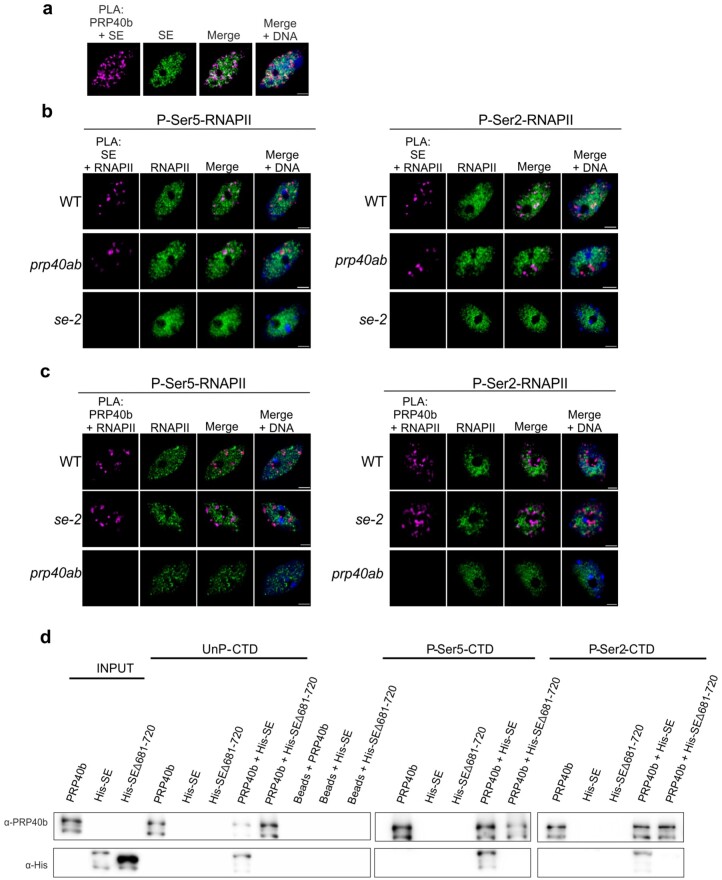
Figure 1.
AtPRP40 regulates the association of SE with RNAPII. A, Close proximity of AtPRP40b and SE in the cell nucleus (first image, magenta signals) analyzed by PLA in view of SE nuclear localization (second image, green signals). DNA was stained with Hoechst (blue). Scale bar = 2.5 µm. B, Close proximity of SE and RNAPII phosphorylated at CTD Ser5 or Ser2 in WT, prp40ab, and se-2 plants (first columns, magenta signals) in view of P-Ser5-RNAPII or P-Ser2-RNAPII nuclear localization (second columns, green signals) detected by PLA. DNA was stained with Hoechst (blue). Scale bar = 2.5 µm. C, Close proximity of AtPRP40b and RNAPII phosphorylated at CTD Ser5 or Ser2 in WT, se-2, and prp40ab plants (first columns, magenta signals) in view of P-Ser5-RNAPII or P-Ser2-RNAPII nuclear localization (second columns, green signals) detected by PLA. DNA was stained with Hoechst (blue). Scale bar = 2.5 µm. D, In vitro pull-down assays using recombinant AtPRP40b and SE proteins (both full length and the shortened C-terminus variant Δ681–720 were used) and biotinylated CTD peptides in unphosphorylated (UnP) or phosphorylated Ser5 (P-Ser5-CTD) or Ser2 (P-Ser2-CTD) forms. The recombinant proteins were incubated with CTD peptides immobilized on beads. The obtained complexes were washed, eluted, and analyzed by immunoblot. Input represents 1/10 of the protein sample.

