Figure 2.
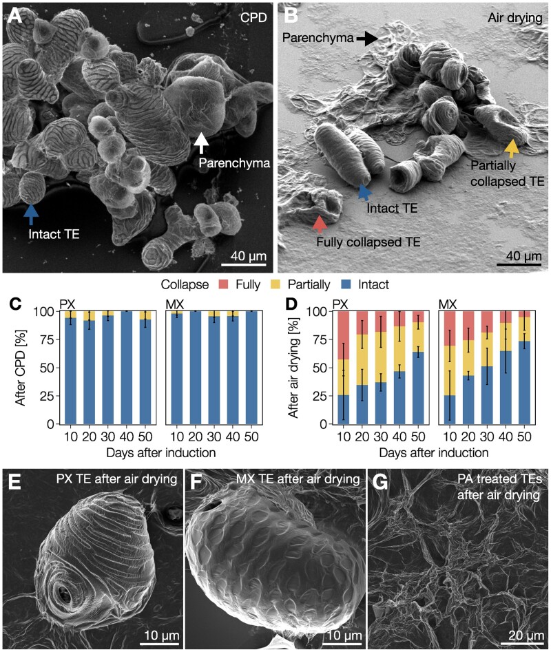
Gradual postmortem lignification enables all TE morphotypes to resist extreme differentials. A, Scanning electron micrograph of 30-d-old isolated TEs and parenchyma cells produced from iPSCs and prepared using CPD. Note that both TEs and parenchyma are intact, as indicated by the blue and white arrows, respectively. B, Scanning electron micrograph of 30-d-old isolated TEs and parenchyma cells produced from iPSCs and prepared using air drying. Note that parenchyma cells (black arrow) are completely flattened whereas TEs were either fully collapsed (red arrow), partially collapsed (yellow arrow), or intact (blue arrow). C and D, Relative proportion of 10- to 50-d-old TEs from iPSCs that were fully collapsed, partially collapsed, or intact after CPD (C) or air drying (D). Error bars represent ± SD of three independent experiments; n = 27–159 individual cells per cell type and time point. E, Scanning electron micrograph of a 30-d-old PX TE after air drying. F, Scanning electron micrograph of a 30-d-old MX TE after air drying. G, Scanning electron micrograph of 30-d-old unlignified TEs treated with PA after air drying.

