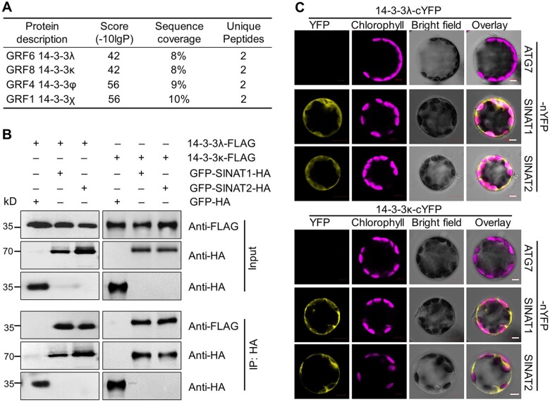
Figure 1.
SINATs interact with 14-3-3λ and 14-3-3κ proteins in vivo. A, Screening for SINAT1-interacting proteins by IP-MS analysis. Total protein was extracted from GFP-SINAT1-HA transgenic seedlings grown on MS medium, immunoprecipitated by anti-GFP agarose beads, and then subjected to in-gel digestion and MS analysis. Transgenic seedlings expressing free GFP-HA were used as a negative control. B, In vivo Co-IP assay validating the association between SINATs (SINAT1 and SINAT2) and 14-3-3s (14-3-3λ and 14-3-3κ). Constructs encoding 14-3-3λ-FLAG or 14-3-3κ-FLAG and GFP-SINAT1-HA or GFP-SINAT2-HA were transiently co-transfected into protoplasts from WT plants (Col-0) and incubated under light conditions for 16 h before immunoprecipitation with HA affinity agarose beads. GFP-HA was co-transfected with 14-3-3λ-FLAG or 14-3-3κ-FLAG as a negative control. Numbers on the left indicate the molecular weight (kDa) of each band. C, BiFC assay of 14-3-3s (14-3-3λ and 14-3-3κ) and SINATs (SINAT1 and SINAT2) in Arabidopsis protoplasts. Constructs encoding the split nYFP fusions SINAT1-nYFP or SINAT2-nYFP and the cYFP fusions 14-3-3λ-cYFP or 14-3-3κ-cYFP were co-transfected in leaf protoplasts and incubated for 16 h under light conditions. The vector pairs 14-3-3λ-cYFP + ATG7-nYFP and 14-3-3κ-cYFP + ATG7-nYFP were co-transfected as negative controls. Confocal micrographs obtained from YFP, chlorophyll autofluorescence, brightfield, and merged images are shown. Scale bars, 5 μm.

