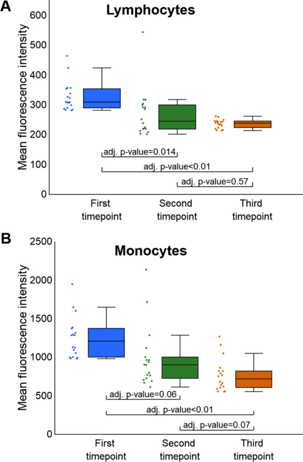
Fig. 1.

Analysis of relative histone acetylation changes in lymphocytes (A) and monocytes (B) between study timepoints. A The change in the fluorescence levels for lymphocytes was statistically significant between the first and both the second and the third timepoints. B In monocytes, we observed a statistically significant decrease in detected fluorescence between the first and the third timepoints. The change was not statistically significant between the first and second timepoints. We did not observe statistically significant differences in the levels of fluorescence intensity between the second and third timepoints in neither lymphocytes (A) nor monocytes (B)
