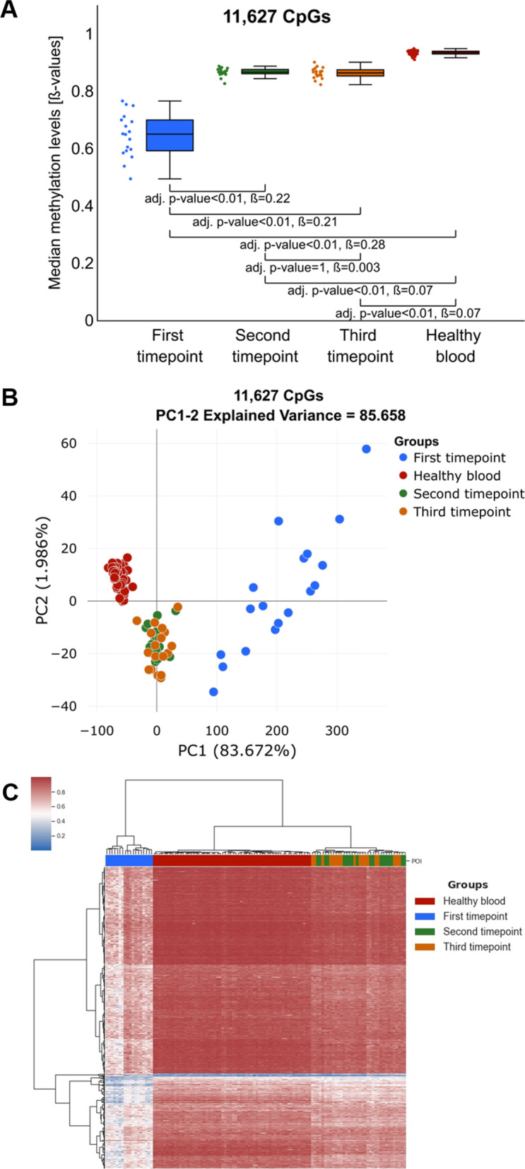
Fig. 3.

Comparison of the methylation changes at a subset of CpG sites that we identify to display diet-related methylation changes. A Comparison of the median methylation levels for analysed groups including: first (blue), second (green), third (orange) timepoints and healthy controls (red). The difference at identified CpGs is significantly larger than the ones observed in Fig. 3B, indicating that methylation at this subset of CpG sites was most affected in the study. B PCA plot based on the identified subset of CpG sites indicates a significant change in the methylation profile of this subset of CpG sites between the first and the second/third timepoints. C Heatmap illustrating unsupervised clustering analyses indicates significant hypomethylation (blue colour in the heatmap) at the first study timepoint and significant increase in the methylation level towards the levels observed in healthy controls (red colour in the heatmap) at the second/third timepoints
