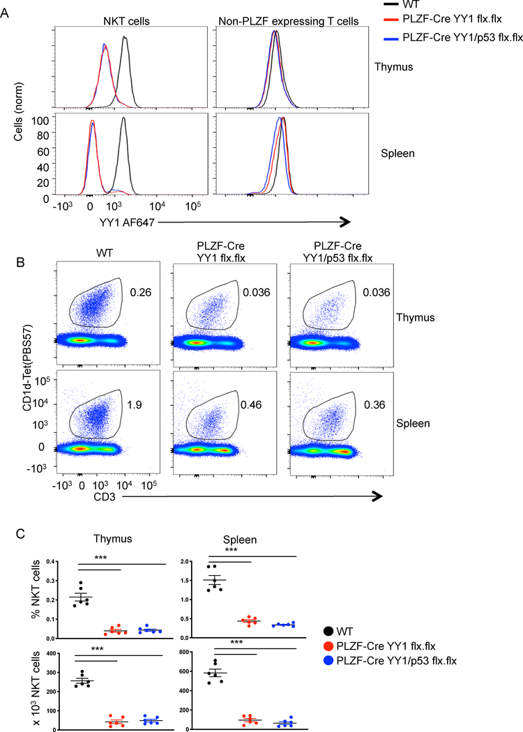
Figure 1. Cell specific deletion of YY1 causes a decrease in the number of NKT cells.
The indicated tissues were collected from C57BL/6 (WT) mice, PLZF-Cre YY1 flx.flx mice or PLZF-Cre YY1/p53 flx.flx mice and analyzed by FACS with the indicated antibodies. (A) YY1 expression was compared between WT NKT cells (MHCII−, CD3+, CD1d tet+, CD24−), PLZF-Cre YY1 flx.flx NKT cells, and PLZF-Cre YY1/p53 flx.flx NKT cells (left) isolated from the thymus (top) or spleen (bottom). YY1 expression was also compared between conventional T cells (MHCII−, CD3+, CD1dtet−, PLZF−) (right) isolated from WT mice, PLZF-Cre YY1 flx.flx mice, and PLZF-Cre YY1/p53 flx.flx mice. Data are representative of 5 or more independent experiments. (B, C) The frequency and absolute numbers of NKT cells (MHCII−, CD3+, CD1dtet+) in the thymus and spleen of WT mice, PLZF-Cre YY1 flx.flx mice, and PLZF-Cre YY1/p53 flx.flx mice were determined by FACS. Representative plots are shown in (B) and pooled data concerning the frequency (C, top) and absolute number (C, bottom) are in (C). FACS plots show representative results from at least 5 mice analyzed in at least 5 independent experiments (A, B). Graphs (C) show compiled data from 6 mice, examined in 5 independent experiments. The horizontal lines indicate the mean (±s.e.m.). ***P<.001 determined by One-Way Anova.

