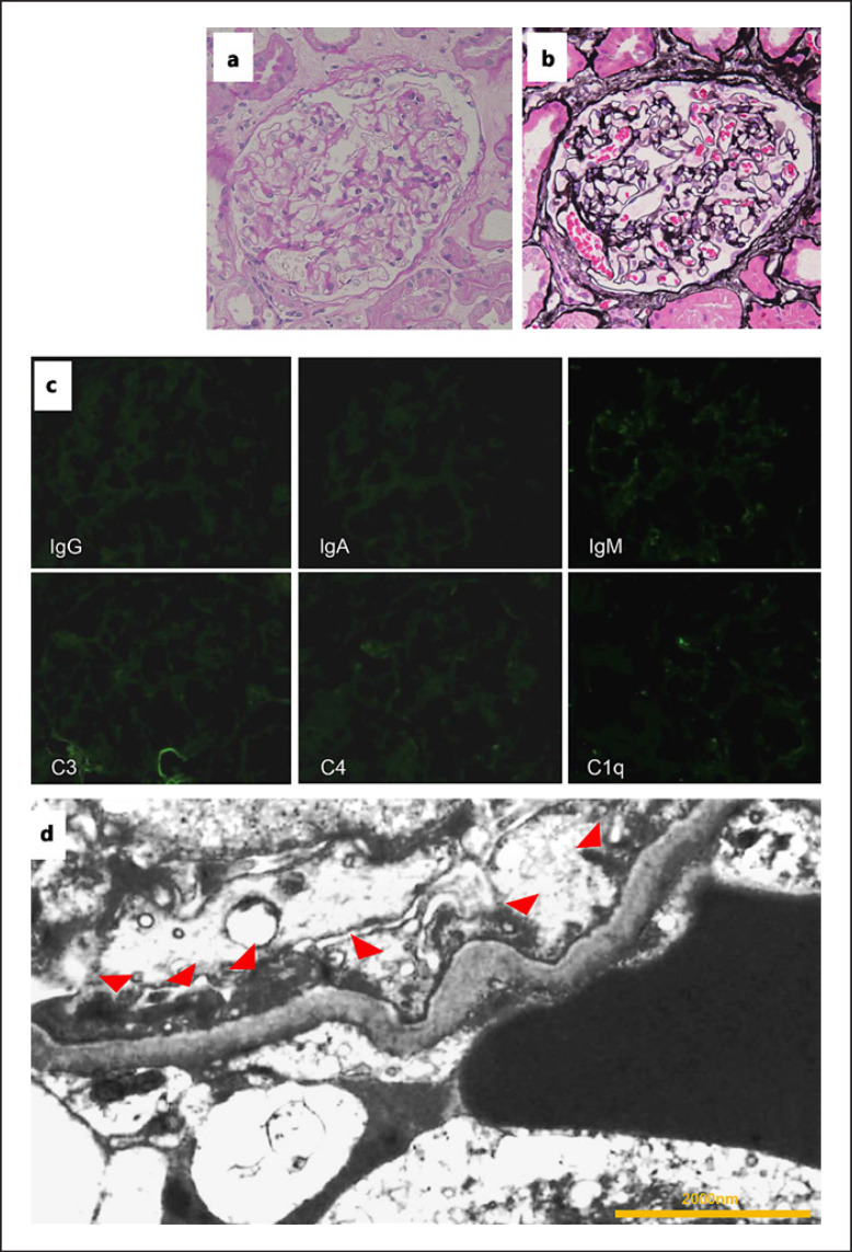
Fig. 1.
Pathological findings. a Optical microscopy reveals no change in the glomeruli (periodic acid-Schiff staining; magnification, ×400). b No changes are observed in the glomerular basement membrane (periodic acid-methenamine silver staining; magnification, ×400). c Immunofluorescence studies show no particular staining. d Electron microscopy shows foot process effacement (red arrow heads). There are no electron dense deposits (magnification, ×7,000).

