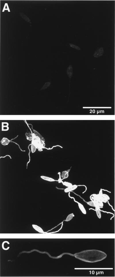
FIG. 4.
Confocal fluorescence microscopy of transfectants for cell surface gp63. Glutaraldehyde-fixed cells were treated first with anti-gp63 antiserum and then with fluorescein isothiocyanate-conjugated secondary antibodies; those treated with normal serum and/or with the secondary antibodies alone served as controls (see Materials and Methods for details). (A) P6.5/1.9R cells viewed at settings to reduce the images to the background level; (B) P6.5/1.9 cells with intensive signals for gp63 viewed under the same settings; (C) surface localization of gp63 in a P6.5/1.9 cell revealed under appropriate settings.

