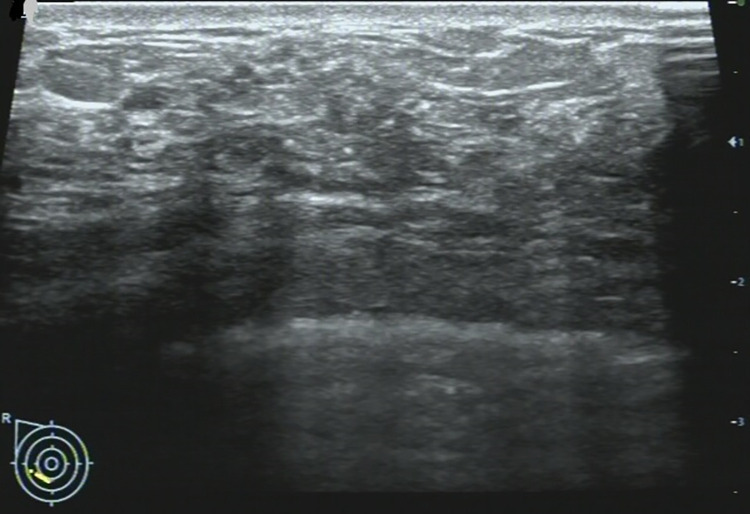
Fig 2. 36-year-old woman with a non-mass lesion in the right breast.
Image shows the breast lesion locates at outer inferior quadrant, shows irregular heterogeneous isoechoic appearance without space-occupying, with scattering fine hyperechoic foci (microcalcifications) in it, and the border and margin is ambiguous. It is pathologically confirmed a non-special invasive breast carcinoma.

