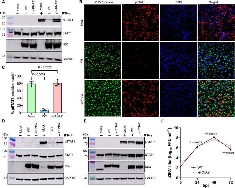
Fig. 7. ZIKV sfRNA inhibits type I and III IFN signaling by suppressing phosphorylation and nuclear translocation of STAT1.
(A) Effect of ZIKV sfRNA on the phosphorylation of STAT1 in response to type I IFN. (B) Immunofluorescent detection of Tyr701-phosphorylated STAT1 in ZIKV-infected Vero cells treated with IFN-α1. The image is representative of three independent experiments that showed similar results. DAPI, 4′,6-diamidino-2-phenylindole. (C) Image quantification for (B). The pSTAT1-positive nuclei were quantified in ZIKV-positive cells in three independent experiments. (D and E) Effect of ZIKV sfRNA on the phosphorylation of STAT1 in response to type II (E) and type III (D) IFN. The band in mock after probing for NS3 in (E) represents nonspecific antibody binding and has different NS3 molecular weight. (F) Replication of WT and sfRNA-deficient ZIKV in STAT1-deficient U3A cells. Cells were infected at MOI = 0.1; titers were determined by a foci-forming assay of C6/36 cells. In (A) to (E), Vero (A to C and E) or HTR-8 cells (D) were infected with WT or xrRNA2′ ZIKV or left uninfected (mock). At 48 hpi, cells were treated with human IFN-α1 (A to C), IFN-λ1 (D), or IFN-γ (E) for 20 (A to C and E) or 60 min (D). Levels of Tyr701-phosphorylated STAT1 (pSTAT1) and total STAT1 indicate phosphorylation and expression of STAT1, respectively. Levels of ZIKV NS3 indicate viral loads in the infected cells. GAPDH levels indicate the total protein input. In (C) and (F), the values are the means from three independent experiments ± SD. The statistical analysis is by Student’s t test in (C) and one-way ANOVA in (F). All tests are two-sided.

