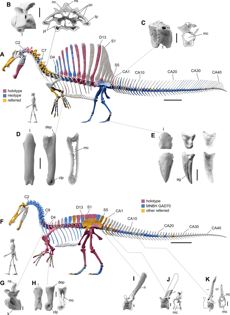
Figure 1. Digital skeletal reconstructions of the African spinosaurids Spinosaurus aegyptiacus and Suchomimus tenerensis.
(A) S. aegyptiacus (early Late Cretaceous, Cenomanian, ca. 95 Ma) showing known bones based on the holotype (BSPG 1912 VIII 19, red), neotype (FSAC-KK 11888, blue), and referred specimens (yellow) and the center of mass (red cross) of the flesh model in bipedal stance (overlap priority: neotype, holotype, referred bones). (B) Cervical 9 (BSPG 2011 I 115) in lateral view and coronal cross-section showing internal air space. (C) Caudal 1 centrum (FSAC-KK 11888) in anterolateral view and coronal CT cross-section. (D) Right manual phalanx I-1 (UCRC PV8) in dorsal, lateral, and sagittal CT cross-sectional views. (E) Pedal phalanges IV-4, IV-ungual (FSAC-KK 11888) in dorsal, lateral, and sagittal CT. (F) S. tenerensis (mid Cretaceous, Aptian-Albian, ca. 110 Ma) showing known bones based on the holotype (MNBH GAD500, red), a partial skeleton (MNBH GAD70, blue), and other referred specimens (yellow) (overlap priority: holotype, MNBH GAD70, referred bones). (G) Dorsal 3 in lateral view (MNBH GAD70). (H) Left manual phalanx I-1 (MNBH GAD503) in dorsal, lateral, and sagittal CT cross-sectional views. (I) Caudal 1 vertebra in lateral view (MNBH GAD71). (J) Caudal ~3 vertebra in lateral view (MNBH GAD85). (K) Caudal ~13 vertebra in lateral view with CT cross-sections (coronal, horizontal) of the hollow centrum and neural spine (MNBH GAD70). ag, attachment groove; C2, 7, 9, cervical vertebra 2, 7, 9; CA1, 10, 20, 30, 40, caudal vertebra 1, 10, 20, 30, 40; clp, collateral ligament pit; D4, 13, dorsal vertebra 4, 13; dip, dorsal intercondylar process; k, keel; mc, medullary cavity; nc, neural canal; ns, neural spine; pc, pneumatic cavity; pl, pleurocoel; r, ridge; S1, 5, sacral vertebra 1, 5. Dashed lines indicate contour of missing bone, arrows indicate plane of CT-sectional views, and scale bars equal 1 m (A, F), 5 cm (B, C), 3 cm (D, E, H–K) with human skeletons 1.8 m tall (A, F).

