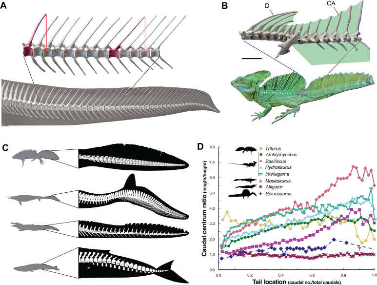
Figure 4. Skeletal comparisons between Spinosaurus aegyptiacus, a basilisk lizard and secondarily aquatic vertebrates.
(A) Tail in S. aegyptiacus showing overlap of individual neural spines (red) with more posterior vertebral segments. (B) Sail structure in the green basilisk (CT-scan enlargement) and in vivo form and coloration of the median head crest and sail (Basiliscus plumifrons FMNH 112993). (C) Structure of the tail fluke in a urodele, mosasaur, crocodilian, and whale. (D) Centrum proportions along the tail in the northern crested newt (Triturus cristatus FMNH 48926), semiaquatic lizards (marine iguana Amblyrhynchus cristatus UF 41558, common basilisk Basiliscus basiliscus UMMZ 121461, Australian water dragon Intellagama lesueurii FMNH 57512, sailfin lizard Hydrosaurus amboinensis KU 314941), an extinct mosasaurid (Mosasaurus sp. UCMP 61221; Lindgren et al., 2013), an alligator (Alligator mississippiensis UF 21461), and Spinosaurus (S. aegyptiacus FSAC-KK 11888). Data in Appendix 2.

