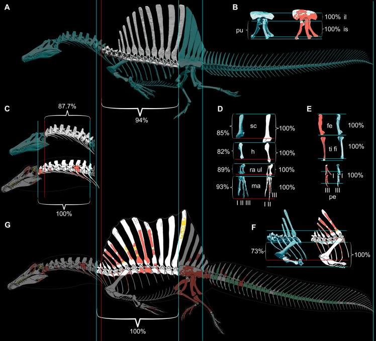
Figure 9. Comparison of skeletal reconstructions for Spinosaurus aegyptiacus in left lateral view.
(A) Digital skeletal reconstruction from this study in left lateral view. (B) Pelvic girdle. (C) Cervical column (C1–10). (D) Pectoral girdle and forelimb. (E) Hind limb. (F) Anterior trunk. (G) Silhouette skeletal drawing from the aquatic hypothesis (from Ibrahim et al., 2020b). On one side of each length comparison, one or two blue lines are shown that register the alternative reconstructions. The opposing end of each length comparison either has a single blue line (when comparisons match, both 100%) or a red line as well for the shorter one (<100%): A blue line on the right or top sides of each comparison is used for registration. The opposing side has a blue line if reconstructions agree on length (100%), or a blue line for the length estimate in this study and a red line for that of the aquatic hypothesis. In all disparate comparisons, the reconstruction in this study is shorter (percentage given). Skeletal reconstructions (A, G) are aligned by the anterior and posterior margins of the ilium and measured to the cervicodorsal junction (C10-D1); the pelvic girdle (B) is aligned along the ventral edge of the sacral centra and base of the neural spines and measured to the distal ends of the pubis and ischium; the cervical column (C) is aligned at the cervicodorsal junction (C10-D1) and measured to the anterior end of the axis (C2); the scapula and components of the forelimb (humerus, ulna, manual digit II, manual phalanx II-1) (D) are aligned at the distal end of the blade and their proximal ends, respectively, and measured to the opposing end of the bone; the components of the hind limb (femur, tibia, pedal digits I, III) (E) are aligned at their proximal ends and measured to the opposing end of the bone; and anterior trunk depth (F) is aligned along the ventral edge of the centrum and neck of the spine of D6 and measured to the ventral edge of the coracoid. II-1, manual phalanx II-1; Ili, ilium; F, femur; H, humerus; Ish, ischium; l, left; Pe, pes; Pu, pubis; Ma, manus; r, right; RaU, radius-ulna; Sc, scapula; TF, tibia-fibula.

