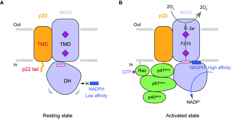
(
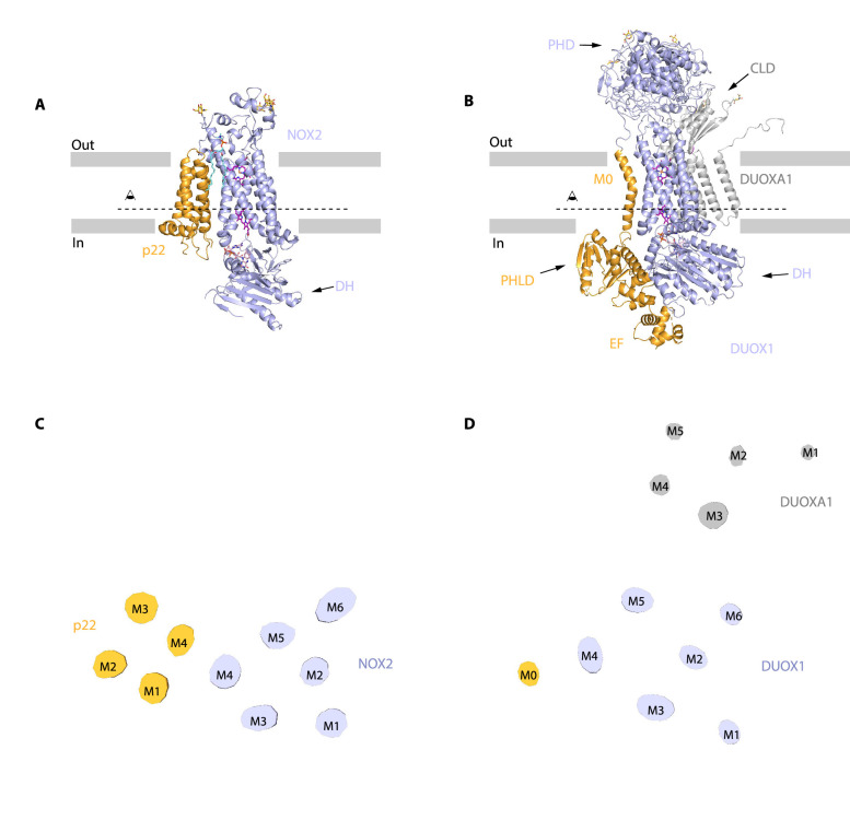
A) Structure of human NOX2 and p22 complex (PDB ID: 8GZ3) in cartoon representation. The colors of each individual domain are the same as in
Figure 1I. The approximate boundaries of the phospholipid bilayer are indicated as gray thick lines. Sugar moieties, haems, FAD, and lipids are shown as gold, purple, pink, and aquamarine sticks, respectively. (
B) Structure of human DUOX1-DUOXA1 complex (PDB ID: 7D3F) in cartoon representation. PHD: peroxidase homology domain of DUOX1; PHLD: pleckstrin homology-like domain of DUOX1; EF: EF-hand calcium-binding module of DUOX1; DH: dehydrogenase domain of DUOX1; CLD: claudin-like domain of DUOXA1. The M0 helix, PHLD, and EF are colored in orange, and the other part of DUOX1 is colored in blue. The DUOXA1 is colored in gray. (
C) Top view of the cross-section of NOX2-p22 cryo-EM map on the transmembrane layer at the position indicated as a dashed line in A. For clarity, the cryo-EM map was low-pass filtered to 7 Å. (
D) Top view of the cross-section of DUOX1-DUOXA1 cryo-EM map on the transmembrane layer at the position indicated as a dashed line in B.


