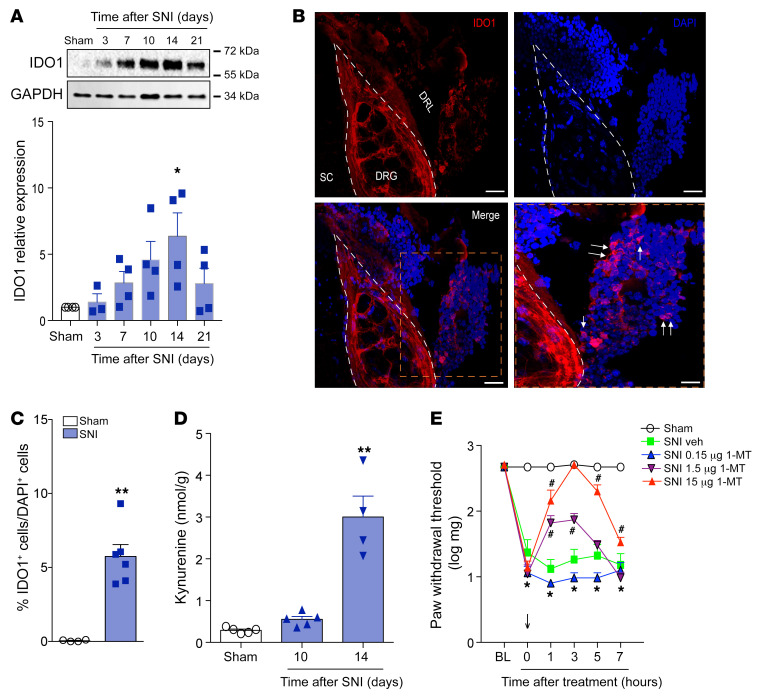
Figure 5. Cells expressing IDO1 accumulate in the DRL after SNI and contribute to the maintenance of neuropathic pain.
(A) Time course of IDO1 expression in the DRGs plus DRL tissues (n = 4 per time point). (B) Representative images showing immunoreactivity for IDO1 (red color) double labeled with DAPI (cell nuclei, blue) in the ipsilateral region containing DRG (L4), DRL, and spinal cord (SC) from SNI mice (14 days after SNI). Scale bars: 100 μm. (C) Quantification of IDO1-expressing cells in the DRL from SNI mice (14 days after SNI) or sham mice (n = 6). (D) Time course of kynurenine levels in the ipsilateral dorsal horn of the spinal cord of mice after sham (14 days) and SNI surgeries (n = 5 per time point). (E) Mechanical nociceptive threshold was determined before and 14 days after SNI. Mice were treated intrathecally with vehicle or 1-methyl-DL-tryptophan (1-MT, 0.15–15 μg/site) and mechanical allodynia was measured up to 7 hours after treatment (n = 5). Data are expressed as mean ± SEM. *P < 0.05, **P < 0.001 versus sham group; #P < 0.05 versus vehicle-treated mice by 1-way ANOVA with Bonferroni’s post hoc test (A and D), unpaired 2-tailed Student’s t test (C), or 2-way ANOVA with Bonferroni’s post hoc test (E).

