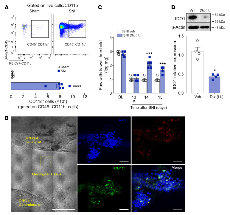
Figure 6. IDO1-expressing DCs accumulate in the DRL after SNI and contribute to the maintenance of neuropathic pain.
(A) Representative dot plots and quantification of CD45+CD11b–CD11c+ cells (DCs) in DRGs plus DRLs harvested 14 days after sham or SNI surgery from WT mice (n = 5). (B) Representative images showing immunoreactivity for IDO1 (red color) double labeled with anti-CD11c (DCs) in the ipsilateral DRLs (L4) from SNI mice (14 days after SNI). Scale bars: 50 μm (left) and 100 μm (right). (C) Mechanical nociceptive threshold was determined before and 13 days after SNI. Mice were treated intrathecally with vehicle or diphtheria toxin (Dtx, 20 ng/site) and mechanical allodynia was measured up to 48 hours after treatment (n = 5). (D) Western blotting analysis of IDO1 expression in the DRGs plus DRL tissues harvested 15 days after SNI mice were treated with vehicle or Dtx (n = 4). Data are expressed as mean ± SEM. *P < 0.05, ***P < 0.001, ****P < 0.0001 versus sham group by unpaired 2-tailed Student’s t test (A and D) or 1-way ANOVA with Bonferroni’s post hoc test (C).

