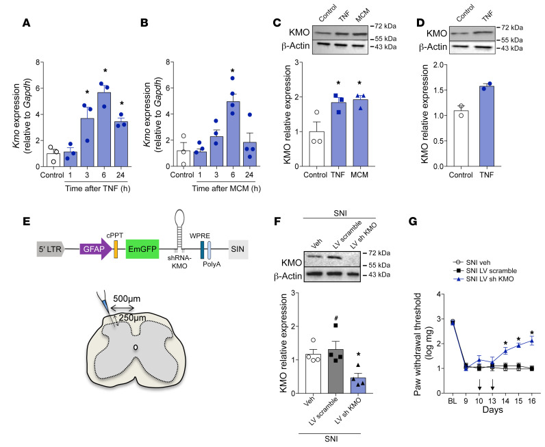
Figure 9. Astrocyte-expressed KMO maintains neuropathic pain.
Primary cultured astrocytes from mouse cortex were stimulated with (A) TNF (10 ng/mL) or (B) microglia-conditioned medium (MCM). After indicated time points, mRNA was extracted and Kmo expression was analyzed by real-time PCR (n = 3–4). (C) Primary cultured astrocytes from mouse cortex or (D) differentiated U87-MG cells were stimulated with TNF (10 ng/mL) or MCM (n = 2–3). The expression of KMO in the cell extract was evaluated 24 hours after stimulation by Western blotting. (E) Schematic of the astrocyte-specific shRNA lentiviral vector (LV) used to knock down KMO in astrocytes of the spinal cord. (F) Mechanical nociceptive threshold was determined before and 9 days after SNI followed by intrathecal treatment with lentiviral vectors expressing shRNA control or shRNA Kmo (n = 4–5) on days 10 and 13 after SNI. (G) Mechanical allodynia was measured up to 16 days after SNI and ipsilateral dorsal horn of the spinal cord was collected for analyses of KMO expression (n = 4). Data are expressed as mean ± SEM. *P < 0.05 versus medium treated; #P < 0.05 versus mice treated with scramble shRNA by 1-way ANOVA with Bonferroni’s post hoc test (A–C and G) or 2-way ANOVA with Bonferroni’s post hoc test (F).

