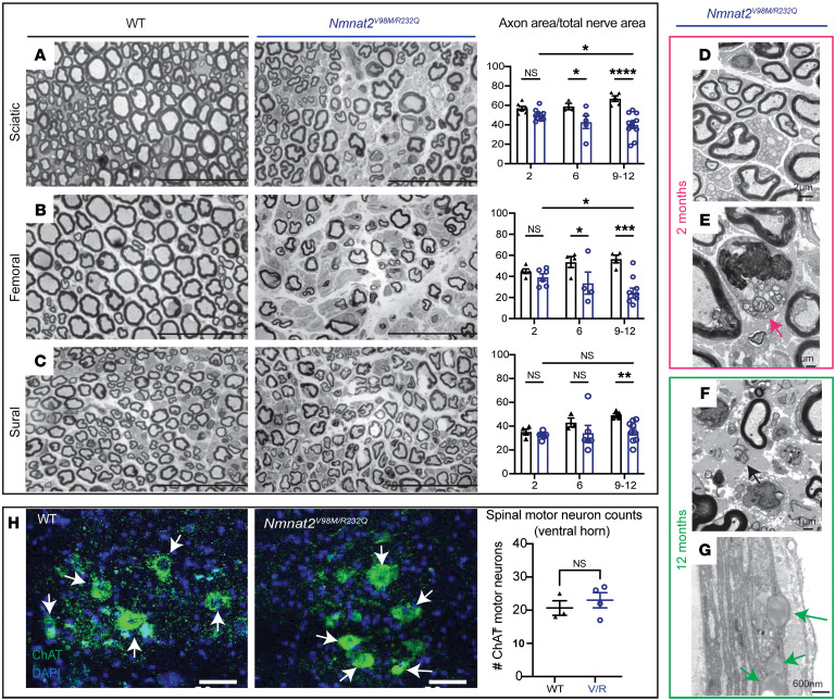
Figure 3. Nmnat2 variants cause progressive axon loss in mice.
(A–C) Representative images of sciatic (A), femoral (B), and sural (C) nerves in 9–12-month-old Nmnat2V98M/R232Q (n = 9) or WT (n = 5) mice. Percent axonal area/total nerve area are indicated to the right (n = 4–11 mice per age cohort, per genotype). Scale bars: 50 μm. (D) Nmnat2V98M/R232Q sciatic nerve (2 months): dense population of large and small myelinated axons with little intervening extracellular space. (E) Nmnat2V98M/R232Q sciatic nerve (2 months): macrophage containing axonal and myelin debris in the endoneurial. (F) Nmnat2V98M/R232Q sciatic nerve (12 months): patches of marked axon loss with increased collagen and wispy processes of SC. Scattered macrophages with axonal and myelin debris were identified. (G) Nmnat2V98M/R232Q sciatic nerve (12 months): presence of large perineurial droplets of neutral fat. (H) Representative images of ChAT immunostaining in 12-month-old Nmnat2V98M/R232Q (n = 4) or WT (n = 3) spinal cord (ventral horn), scale bars: 50 μm. Quantification of number of ChAT+ motor neuron cell bodies in the ventral horn to the right. All data are presented as mean ± SEM. Statistical significance determined by Student’s unpaired, 2-tailed t test or 2-way ANOVA with multiple comparisons. *P < 0.05, **P < 0.01, ***P < 0.001, ****P < 0.0001.

