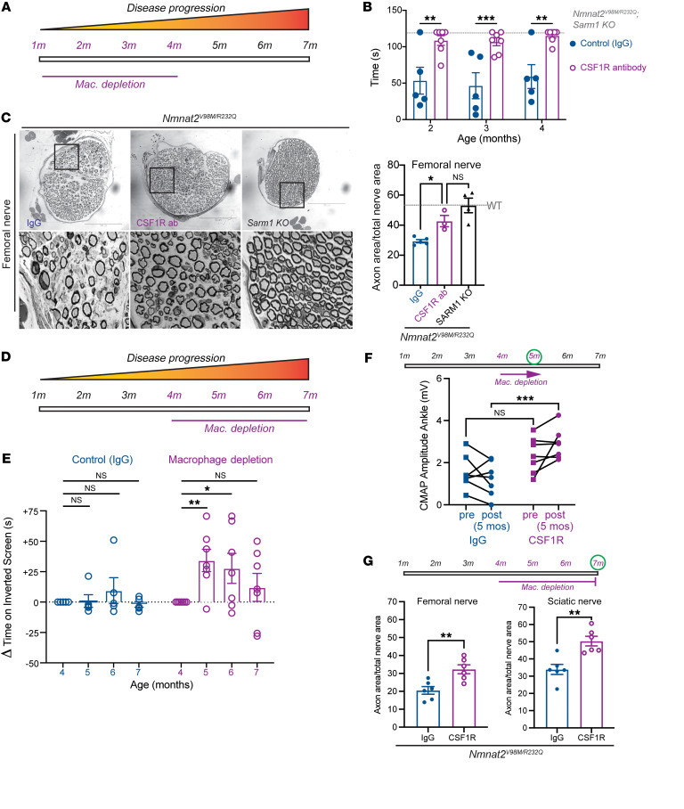
Figure 8. Macrophage depletion rescues motor defects and axon loss in Nmnat2V98M/R232Q mice.
(A) Schematic of CSF1R antibody-mediated macrophage depletion in young (beginning at 1-month-old) Nmnat2V98M/R232Q mice. (B) Average time suspended from an inverted screen (max. 120 seconds) for IgG control (n = 5) and CSF1R-treated Nmnat2V98M/R232Q (n = 7) mice. Statistical significance was determined by 2-way ANOVA with multiple comparisons. (C) Representative images of femoral nerve from IgG (control) (n = 5), CSF1R (n = 3), or Sarm1-KO Nmnat2V98M/R232Q (n = 4) mice at 4 months. Scale bars: 150 μm. Percent axonal area/total nerve area for femoral nerve calculated to the right. (D) Schematic of CSF1R antibody-mediated macrophage depletion in aged (beginning at 4-month-old Nmnat2V98M/R232Q mice. (E) Change in inverted screen time (s) from pre-treatment (4 months) measured at 5, 6, and months, comparing CSF1R treatment (n = 7) and IgG (control) (n = 5). Statistical significance was determined by 2-way ANOVA with multiple comparisons. (F) CMAP amplitude (ankle) of Nmnat2V98M/R232Q mice before and after 1 month of CSF1R treatment (n = 7) or IgG treatment (n = 5). (G) Percent axonal area/total nerve area for femoral nerve and sciatic nerve calculated at 7 months (3 months of macrophage depletion (n = 6) or treatment with isotype control IgG (n = 6). All data are presented as mean ± SEM. Statistical significance determined by Student’s unpaired t test. *P < 0.05, **P < 0.01, ***P < 0.001.

