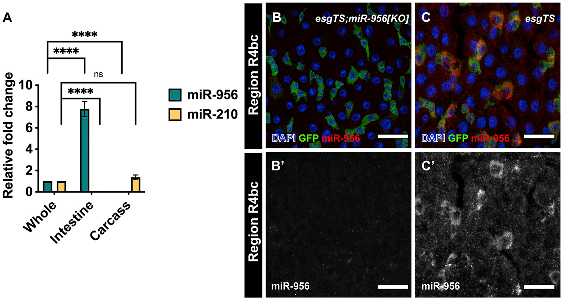
Figure 1. miR-956 is enriched in Drosophila intestinal tissue.

(A) qPCR analysis of miR-956 and miR-210 levels in intestinal tissues and carcass relative to whole animals. For each experiment, samples were collected from three separate animals in triplicates. Statistical significance of the difference in miRNA levels in intestinal and carcass samples relative to whole tissue is indicated. (B and C) esgTS-labeled progenitor cells (green) in (B) esgTS; miR-956[KO] mutants, which were used as a control, and (C) wild-type esgTS animals, with counterstaining of all cell nuclei (DAPI in blue) and miR-956 using RNA in situ probes (red). (B′–C′) Grayscale images of miR-956 RNA in situ. Scale bar, 25 μm. Data shown as mean ± SEM. Significance values: ns, not significant; ****p < 0.0001.
