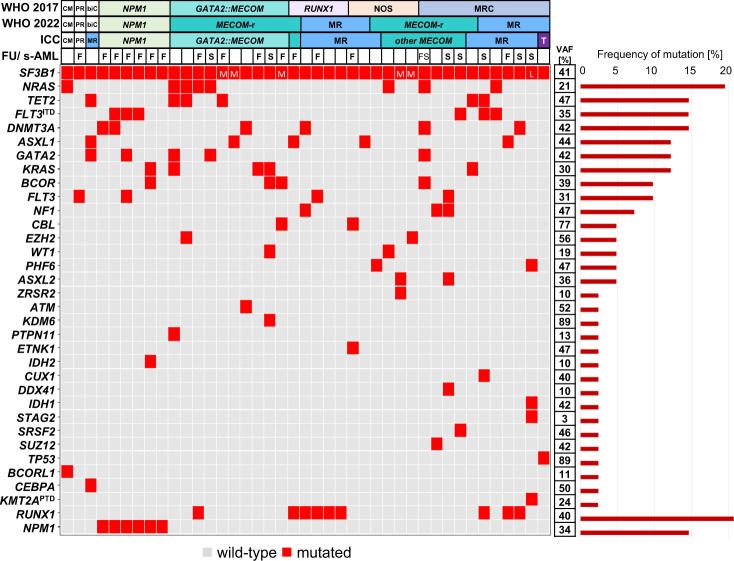
Fig. 2. Molecular characterization of AML patients with mutated SF3B1.
Illustration of all 41 samples, each column represents one patient. Genes (gray: wild-type; red: mutated) as well as the WHO and ICC entities are given for each patient. Secondary AMLs (s-AMLs) are marked with “S” and those with available follow-up (FU) data with “F”. VAF variant allelic frequency (mean), CM CBFB::MYH11, PR PML::RARA, biC biallelic CEBPA, NOS not otherwise specified, MR(C) myelodysplasia-related (changes), MECOM-r MECOM rearrangement, T mutated TP53, L low VAF (0–14%), M medium VAF (15–29%); Remaining cases showed SF3B1 VAFs ≥30%.

