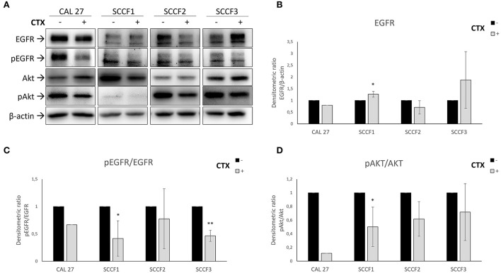
Figure 1.
Inhibition of EGFR pathway by Cetuximab in feline oral squamous cell carcinoma cell lines (SCCF1, SCCF2, SCCF3). (A) Cells were incubated with Cetuximab (CTX) at 100 μg/mL for 12 h and analyzed by western blotting (WB) for EGFR, phospho-EGFR (pEGFR), Akt and phospho-Akt (pAkt). Cetuximab-sensitive CAL 27 cells were included as control. The treatment (+) induced reduction of pEGFR levels in all cell lines with respect to untreated control (–), along with decreased EGFR in SCCF2 and its accumulation in SCCF3 and SCCF1. Impairment of EGFR by Cetuximab was accompanied by a decrease of pAkt levels. WB for β-actin antibody ensured comparable protein loading and allowed normalization. (B) Densitometric analysis of EGFR expressed as densitometric ratio with β-actin. (C,D) Densitometric analysis of pEGFR and pAkt expressed as densitometric ratio pEGFR/EGFR and pAkt/Akt, respectively. Standard deviations are from three repeated, independent experiments (*: statistically significant by t-test, P ≤ 0.05; **: statistically significant by t-test, P ≤ 0.01).

