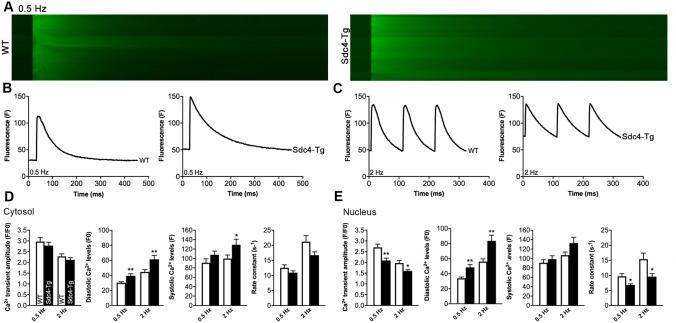
Fig. 4.
Cardiomyocytes from adult Sdc4-Tg mice show increased diastolic Ca2+ levels. A Representative fluorescence confocal microscopy images of cardiomyocytes isolated from adult Sdc4-Tg and wild-type (WT) mice, loaded with the fluorescence-labelled Ca2+ indicator Fluo 4-AM. Representative tracings of cells paced at 0.5 Hz (baseline condition; B) and 2 Hz (stressed condition; C). D, E Ca2+ transient characteristics of n = 16–19 cells from n = 3 WT and Sdc4-Tg mice, paced at 0.5 Hz and 2 Hz. Data are mean ± SEM. Statistical differences were tested using an unpaired t-test vs. respective WT control, *p < 0.05, **p < 0.01

