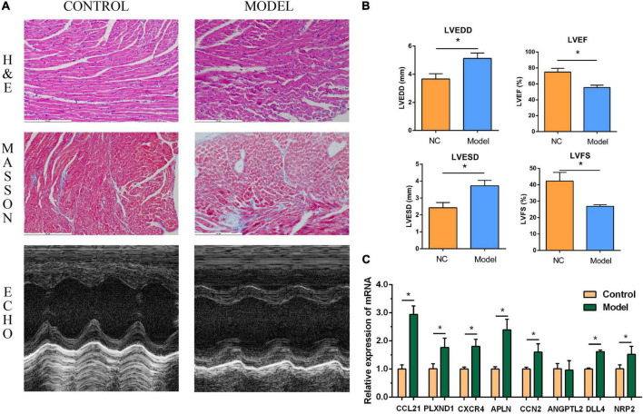
FIGURE 8.
Myocardial injury and impaired heart function were assessed in Type 1 diabetic heart failure mice model. (A) Representative pictures of HE staining, Masson staining, and echocardiography (magnification, 400 ×). (B) Comparison of LVEDS, LVEDD, LVEF, and LVFS between the control group and the model group. (C) Relative mRNA expression of each core gene in control group and model group. *P < 0.05.

