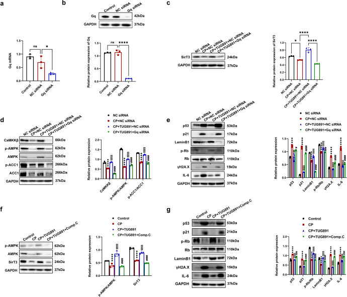
Fig. 9.
Activation of FFAR4 reversed the Sirt3 expression via Gq/CaMKKβ/AMPK signaling in cisplatin-stimulated TCMK-1 cells. a, b Relative mRNA and protein expression of Gq in TCMK-1 cells transfected with Gq siRNA (n = 3; *P < 0.05, ****P < 0.0001, ns no significant difference). c Protein expression of SirT3 in TCMK-1 cells transfected with Gq siRNA detected by western blotting and quantified by densitometry (n = 3; ****P < 0.0001, *P < 0.05). d Protein expression of CaMKKβ, p-AMPK/AMPK, and p-ACC1/ACC1 in TCMK-1 cells transfected with Gq siRNA detected by western blotting and quantified by densitometry (n = 3; ****P < 0.0001, CP + NC siRNA vs. NC siRNA; $$$$P < 0.0001, CP + TUG891 + NC siRNA vs. CP + NC siRNA; ####P < 0.0001, CP + TUG891 + Gq siRNA vs. CP + TUG891 + NC siRNA). e Protein expression of p53, p21, p-Rb/Rb, LaminB1, ɣH2A.X and IL-6 in TCMK-1 cells transfected with Gq siRNA detected by western blotting and quantified by densitometry (n = 3; ****P < 0.0001, ***P < 0.001, CP + NC siRNA vs. NC siRNA; $$$$P < 0.0001, $$P < 0.01, CP + TUG891 + NC siRNA vs. CP + NC siRNA; ####P < 0.0001, #P < 0.05, CP + TUG891 + Gq siRNA vs. CP + TUG891 + NC siRNA). f TCMK-1 cells were preincubated with 10 μM compound C and TUG891 for 1 h, followed by cisplatin for 6 h. Protein expression of p-AMPK/AMPK and SirT3 detected by western blotting and quantified by densitometry (n = 3; ****P < 0.0001, CP vs. Control; $$$$P < 0.0001, CP + TUG891 vs. CP; ####P < 0.0001, CP + TUG891 + Comp.C vs. CP + TUG891). g Protein expression of p53, p21, p-Rb/Rb, LaminB1, ɣH2A.X and IL-6 detected by western blotting and quantified by densitometry (n = 3; ****P < 0.0001, CP vs. Control; $$$$P < 0.0001, CP + TUG891 vs. CP; ####P < 0.0001, CP + TUG891 + Comp.C vs. CP + TUG891). Data are presented as mean ± SD. CP cisplatin

