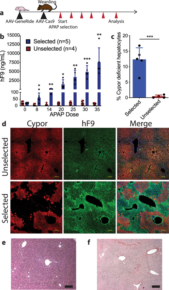
Fig. 2. Expansion of hepatocytes with integration of GR-Cypor scgRNA by acetaminophen.

a Schematic of the experimental timeline. The GR-Cypor scgRNA rAAV vector containing a hF9 transgene and Cypor scgRNA with arms of homology to the Albumin locus was delivered to neonatal WT mice. At weaning, the mice were given rAAV-spCas9 and biweekly APAP administration was started. Mice were bled for hF9 concentration following every 5th dose of APAP administered. b hF9 concentrations in mice that were selected with APAP compared to unselected mice. Biologically independent mice were analyzed. Data are presented as mean values and error bars represent standard deviation (Dose 8 p = 0.015; Dose14 p = 0.008; Dose 20 p = 0.039; Dose 25 p = 0.0051; Dose 30 p = 0.0006; Dose 35 p = 0.006). c Quantification of Cypor deficient hepatocytes by indel frequency following terminal harvest. Biologically independent mice were analyzed. Data are presented as mean values and error bars represent standard deviation (p = 0.0005). Selected n = 5 animals; No selection n = 4 animals. d Representative immunofluorescent staining against Cypor and hF9 in the liver of unselected mice (no APAP treatment) and selected mice (received APAP treatment) at terminal harvest. Scale bars represent 200 µm. e Hematoxylin and eosin staining of a liver section from an APAP selected mouse. Scale bars represent 100 µm. n = 5 mice. f Picro Sirius red staining in an APAP selected mouse. The presence of red staining fibers would indicate fibrosis within the liver. Scale bars represent 100 µm. n = 5 mice. Statistics were calculated using a two-sided unpaired t test. *P < 0.05, **P < 0.01 and ***P < 0.001. Source data are provided as a Source Data file.
