
Fig. 3.
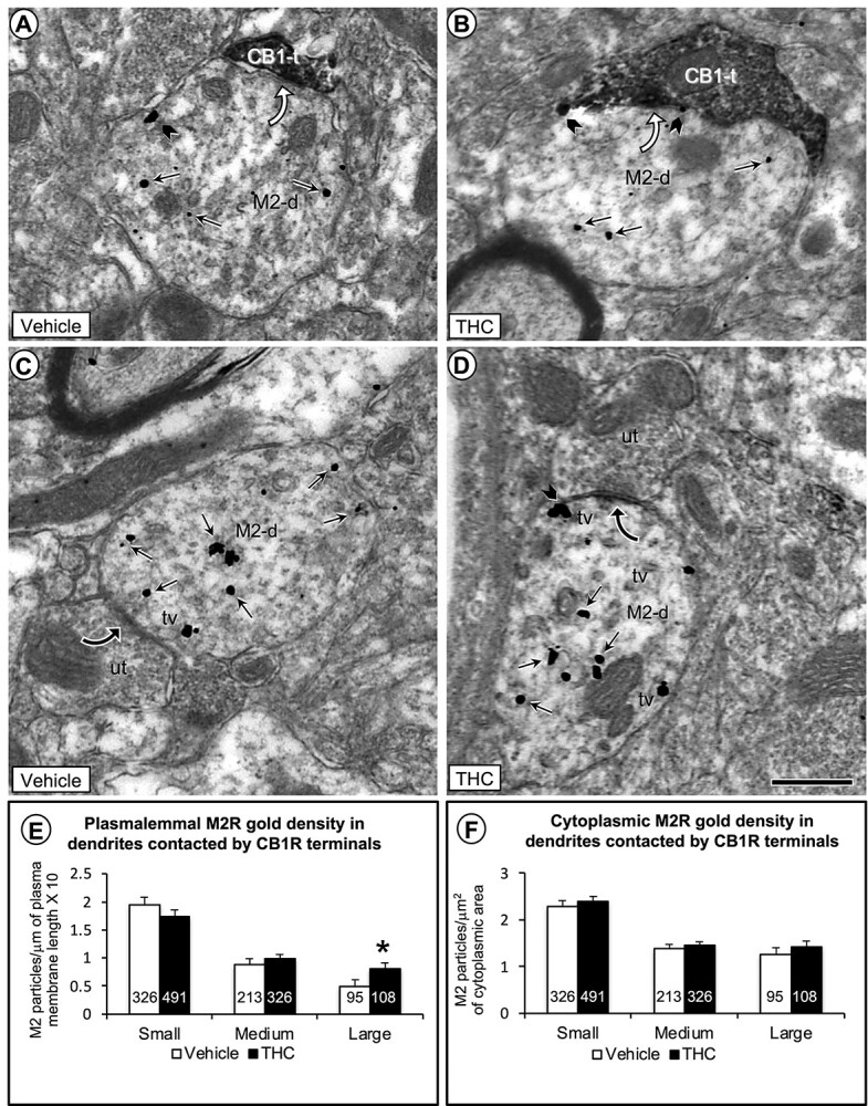
Electron microscopic data comparing the density of M2R immunogold in plasmalemmal and cytoplasmic compartments of large dendritic shafts contacted by CB1R-labeled and unlabeled axon terminals in the PL-PFC of adult mice that received vehicle or THC during adolescence. In a–d), small straight arrows show cytoplasmic and black block arrows plasmalemmal M2R-immunogold particles in large dendrites (M2-d). a and b) As compared to vehicle, the THC-pretreated mice show a qualitative increase in number of M2R immunogold particles on the plasma membrane of large dendrites contacted (curved white arrows) by axon terminals containing CB1R-immunoperoxidase labeling. c and d) In both vehicle and THC-pretreated mice, many M2R-immunogold particles are localized to tubulovesicles (tv) just below the plasma membrane in large dendrites receiving symmetric synapses (curved black arrows) from unlabeled axon terminals. The subsurface vesicles and M2R-immunogold particles are most evident in mice receiving THC compared to vehicle. e) Bar graphs showing a significant increase in the plasmalemmal density of M2R immunogold particles exclusively on the plasma membrane of large dendritic profiles in THC compared with vehicle-injected mice. f) Bar graphs showing no significant differences between vehicle and THC-recipient mice in cytoplasmic density of M2R-immunogold particles in large dendrites. Numbers inside the bars in E-F depict absolute numbers of M2R-immunolabeled dendrites for each category. Asterisk = P < 0.05 ANOVA. Scale bar = 500 nm.