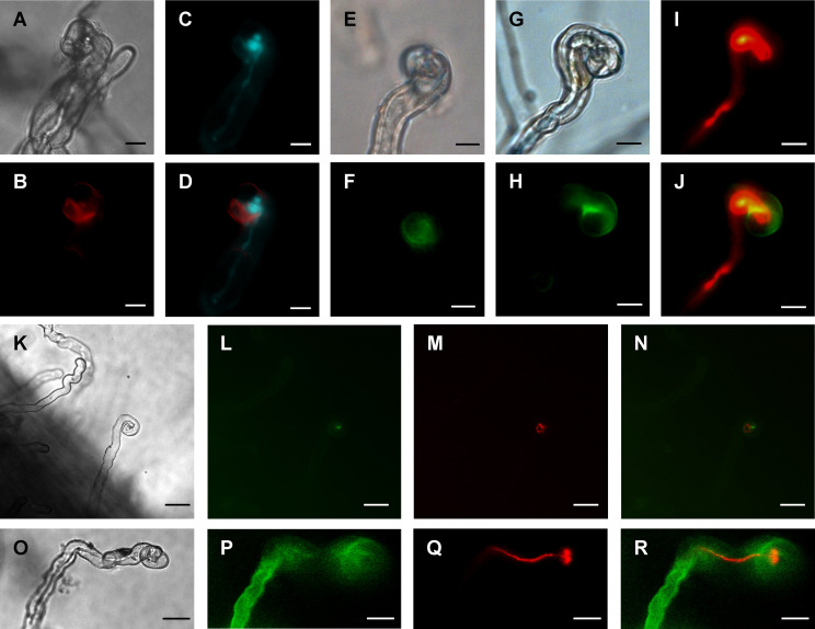
Figure 4.
Subcellular localization of fluorescent MtCHIT5b in root hairs of M. truncatula. (A-D) Transgenic roots of R108 plants expressing MtCHIT5b:mCherry driven by a Ubiquitin gene promoter from L. japonicus were inoculated with S. meliloti Rm41-mTFP1. Root hairs were microscopically analyzed under bright field conditions (A), for red fluorescence emission to detect the MtCHIT5b:mCherry fusion protein (B), and for emission of teal fluorescent signals to visualize Rm41-mTFP1 bacteria (C). The overlay image (D) was obtained from (B, C) and indicates partial co-localization of MtCHIT5b:mCherry with teal fluorescent rhizobia in the infection pocket. (E-N) Transgenic roots expressing MtCHIT5b:GFP driven by the MtCHIT5b native promoter were inoculated with S. meliloti Rm41 (E, F) or S. meliloti Rm41-mCherry (G-N). Root hairs were analyzed under bright field conditions (E, G, K), for green fluorescent signals emitted by MtCHIT5b:GFP (F, H, L), and for red fluorescent signals emitted by Rm41-mCherry (I, M). The overlay images (J, N) indicate co-localization of MtCHIT5b:GFP with red fluorescent rhizobia in the infection pocket (J derived from H and I; N from L and M). (O-R) Similar analysis was performed for transgenic control roots expressing GFP driven by the CaMV 35S promoter. Roots were inoculated with S. meliloti Rm41-mCherry and root hairs were analyzed under bright field conditions (O) and for emission of green (P) or red (Q) fluorescent signals. The overlay image (R) was obtained from (P, Q). Bars = 10 μm in (A-J) and 25 μm in (K-R).

