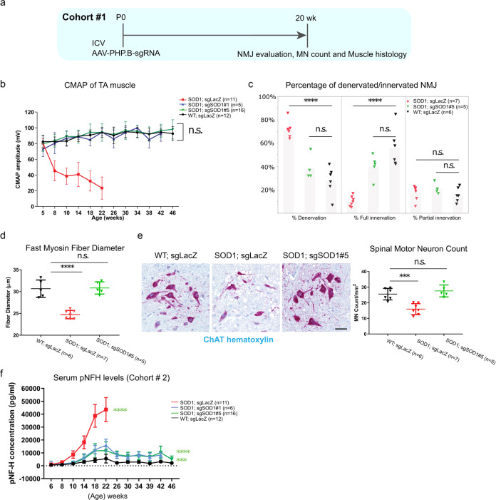
Fig. 3. Neonatal ICV injection of AAV-sgSOD1 prevents NMJ loss, muscle atrophy and neurodegeneration in H11Cas9 SOD1G93A mice.
a Study design of histological and biochemical analysis on H11Cas9 SOD1G93A mice treated with AAV-sgSOD1 (Cohort #1). b CMAP amplitude over disease progression (time). Two-way ANOVA is performed with all groups compared to WT group. c Percentage of fully innervated, denervated and partially innervated NMJs in male mice at 20 weeks of age. d Diameter of fast myosin muscle fiber of tibialis muscles at 20 weeks of age. e Left, representative images of spinal motor neurons stained by ChAT at 20 weeks of age. (scale bar, 50 µm); Right, spinal motor neuron numbers at 20 weeks of age. c–e One-way ANOVA Dunnett’s multiple comparisons test was performed with all groups compared to WT group. f Serum pNFH levels over disease progression (time). Two-way ANOVA was performed, all groups compared to WT group. b–f Data are presented as mean with standard deviation (SD) unless otherwise stated. *p < 0.05, **p < 0.01, ***p < 0.001, ****p < 0.0001, and n.s. not significant. Data was from collected from Cohort #3 in B-E and from Cohort #1 in F.

