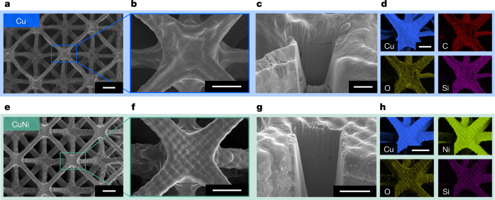
Fig. 2. Morphology of Cu and CuNi microlattices.
a–c,e–g, SEM images of Cu (a–c) and CuNi (e–g) octet lattices, showing multiple unit cells from the top (a,e), a single node (b,f) and a FIB-milled cross-section showing the internal structure of a node from 52° tilt (c,g). d,h, EDS elemental mapping, showing uniform distribution of Cu (d) and uniform distribution of Cu and Ni (h). Scale bars: a,e, 100 µm; b,f, 50 µm; c,g, 20 µm; d,h, 50 µm.

