. 2022 Aug 2;305(3):666–671. doi: 10.1148/radiol.211721

Figure 1:

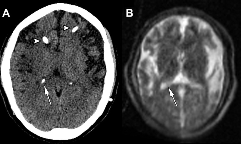
Images in a 42-year-old woman admitted for headaches with a history of congenital hydrocephalus after ventriculoperitoneal shunt placement. (A) Axial head CT scan without contrast shows no significant hydrocephalus at the level of the lateral ventricle atrium (arrow) and partially visualized ventriculostomy catheters (arrowheads). (B) Point-of-care brain MRI scan without contrast obtained with T2-weighted sequence 1 hour after axial head CT and on a similar level shows nonenlarged lateral ventricles at the level of the lateral ventricle atrium (arrow), and the ventriculostomy catheters are more difficult to visualize.

Images in a 42-year-old woman admitted for headaches with a history of congenital hydrocephalus after ventriculoperitoneal shunt placement. (A) Axial head CT scan without contrast shows no significant hydrocephalus at the level of the lateral ventricle atrium (arrow) and partially visualized ventriculostomy catheters (arrowheads). (B) Point-of-care brain MRI scan without contrast obtained with T2-weighted sequence 1 hour after axial head CT and on a similar level shows nonenlarged lateral ventricles at the level of the lateral ventricle atrium (arrow), and the ventriculostomy catheters are more difficult to visualize.