. 2022 Aug 2;305(3):666–671. doi: 10.1148/radiol.211721

Figure 2:

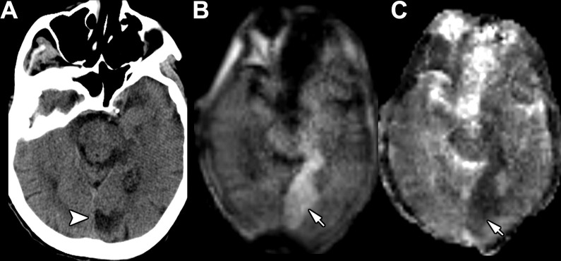
Images in an 84-year-old woman admitted for new facial droop with a history of partial left posterior cerebral artery territory infarction. (A) Axial head CT scan without contrast shows a chronic left posterior cerebral artery infarction (arrowhead). (B, C) Point-of-care brain MRI scans without contrast obtained in the axial plane 1 day later with (B) diffusion-weighted imaging and (C) apparent diffusion coefficient imaging show an acute left posterior cerebral artery territory infarction (arrows) anterior to the area of chronic infarction demonstrated on earlier head CT scans.

Images in an 84-year-old woman admitted for new facial droop with a history of partial left posterior cerebral artery territory infarction. (A) Axial head CT scan without contrast shows a chronic left posterior cerebral artery infarction (arrowhead). (B, C) Point-of-care brain MRI scans without contrast obtained in the axial plane 1 day later with (B) diffusion-weighted imaging and (C) apparent diffusion coefficient imaging show an acute left posterior cerebral artery territory infarction (arrows) anterior to the area of chronic infarction demonstrated on earlier head CT scans.