. 2022 Aug 2;305(3):666–671. doi: 10.1148/radiol.211721

Figure 4:

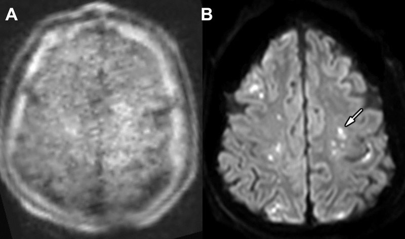
Images in an 89-year-old woman admitted for multiple injuries following a motor vehicle collision. (A) Point-of-care brain MRI scan without contrast obtained in the axial plane with diffusion-weighted imaging shows no significant abnormality. (B) Brain MRI scan without contrast obtained in the axial plane 1 day later with a fixed 3-T scanner and diffusion-weighted imaging shows punctate acute infarctions involving the bilateral centrum semiovale (arrow, left-sided involvement).

Images in an 89-year-old woman admitted for multiple injuries following a motor vehicle collision. (A) Point-of-care brain MRI scan without contrast obtained in the axial plane with diffusion-weighted imaging shows no significant abnormality. (B) Brain MRI scan without contrast obtained in the axial plane 1 day later with a fixed 3-T scanner and diffusion-weighted imaging shows punctate acute infarctions involving the bilateral centrum semiovale (arrow, left-sided involvement).