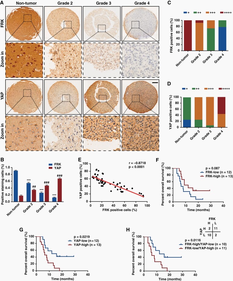
Fig. 1.
The expression level of FRK is negatively correlated with that of YAP in gliomas. (A&B) Representative images of immunohistochemical staining (A) and positive cell percentage (B) of FRK and YAP in nontumor brain tissues (n = 5) and human glioma specimens (n = 45). Scale bar: 20 μm. (C&D) Immunohistochemical scores (+: 0–15%; ++: 15%–30%; +++: 30%–65%; ++++: >65%) of FRK (C) and YAP (D) in gliomas with different grades. (E) The positive staining percentage of FRK and YAP exhibited negative correlation among different specimens (n = 50, r = −0.8718, P < 0.0001). (F–H) Kaplan–Meier survival analysis of high-grade glioma patients expressing indicated proteins (n = 25). The cut-off to define high or low expression was the median value. #, *P < 0.05, ##, **P < 0.01, ###, ***P < 0.001.

