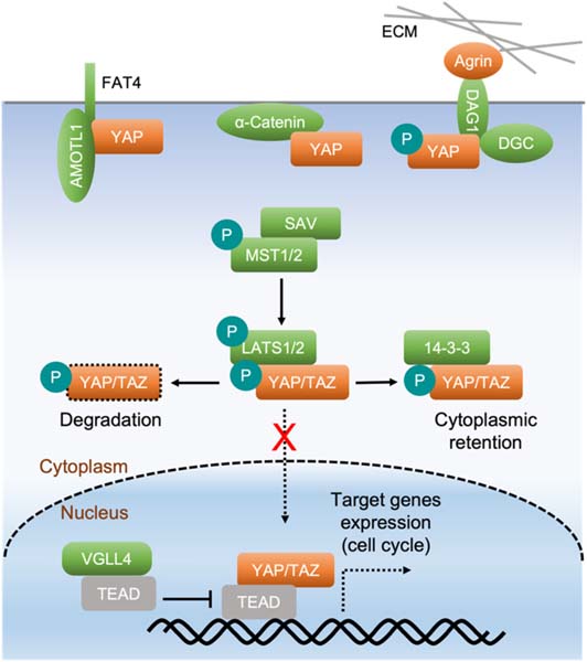
Fig. 1.
The Hippo pathway regulates cardiomyocyte proliferation. The Hippo pathway is composed of several core components: MST1/2, SAV, and LATS1/2. Kinases MST1/2 interact with their adaptor SAV and phosphorylate and activate kinases LATS1/2 that further interact with and phosphorylate YAP and its analog TAZ. Phosphorylated YAP/TAZ are bound and sequestered by 14–3–3 in the cytoplasm or undergo ubiquitination and degradation. When the Hippo pathway is inactivated, unphosphorylated YAP/TAZ translocate into the nucleus, where they interact with their binding partners, such as TEADs, to regulate the expression of downstream target genes that promote cardiomyocyte proliferation. YAP directly interacts with α-catenin, the FAT4 and AMOTL1 complex, and the dystrophin glycoprotein complex (DGC). This causes YAP cytoplasmic retention at the cell membrane, which regulates YAP activity through a Hippo-independent mechanism. The extracellular matrix (ECM) component Agrin physically interacts with the DGC (DAG1 is a protein of the DGC) and disrupts the YAP-DGC interaction, leading to YAP activation.

