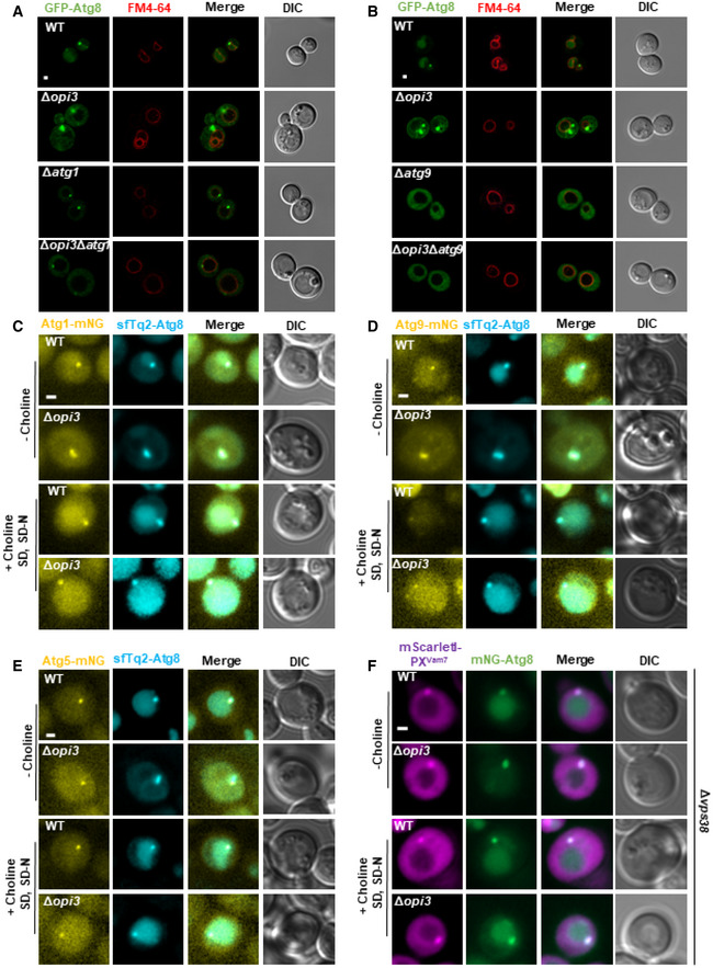
Figure 5. Phospholipid imbalances leads to accumulation of bona fide autophagic structures.

-
A, BRepresentative images of GFP‐Atg8 (green) and vacuole (red, stained with FM4‐64). WT, Δopi3, Δatg1 and Δopi3Δatg1 (A) or Δatg9 and Δopi3Δatg9 (B) cells, all expressing GFP‐Atg8, were grown to log phase in SD‐URA medium, shifted to SD‐N for 3 h and observed by confocal microscopy. Scale bar 1 μm.
-
C–ERepresentative images of mNG‐tagged Atg1 (C), Atg9 (D), or Atg5 (E) colocalized with sfTq2‐Atg8. WT and Δopi3 cells were grown to log phase in SD medium, and shifted to SD‐N. Choline (1 mM) was excluded (−choline) or supplemented during growth and starvation (+choline SD, SD‐N) as indicated. Images were taken during SD‐N (1–3 h) by widefield microscopy. Scale bar 1 μm.
-
FRepresentative images of mNG‐tagged Atg8 colocalized with mScarletI‐PXVam7. WT and Δopi3 cells, expressing mScarletI‐PXVam7 under the CUP1 promoter from the knocked‐out VPS38 locus, were grown to log phase in SD medium in the presence of 10 μM copper sulfate, and shifted to SD‐N in the presence of 10 μM copper sulfate. Choline (1 mM) was excluded (−choline) or supplemented during growth and starvation (+choline SD, SD‐N) as indicated. Images were taken during SD‐N (1–3 h) by widefield microscopy. Scale bar 1 μm.
