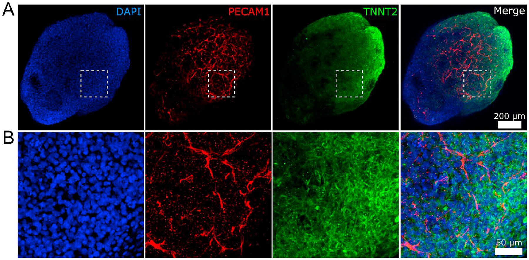
Fig. 5.

Immunostaining and confocal imaging of a representative hHO. (A) Overall view of the hHO. (B) Zoom-in view of the box in panel A. The hHO was fixed on Day 30 and stained with endothelial marker PECAM1 (red), cardiomyocyte marker TNNT2 (green), and nuclei marker DAPI (blue). PECAM1-positive structures reveal formation of the vascular network throughout the hHO. Figures were generated via max projection of z-stacks acquired with the confocal microscope. Scale bars: (A) 200 μm and (B) 50 μm.
