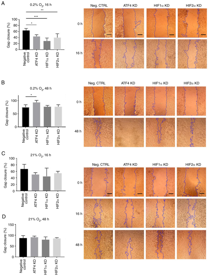
Figure 6.
ATF4, HIF1α and HIF2α KD decrease cell migration in acute hypoxia, but ATF4 KD in chronic hypoxia increases cell migration. (A-D) PANC-1 cells were transiently transfected, scratched and incubated in hypoxia for (A) 16 or (B) 48 h or in normoxia for (C) 16 or (D) 48 h. The negative control were PANC-1 cells exposed to 0.2% oxygen with transfection reagent but no KD small interfering RNA. Images were captured by light microscope at ×50 magnification. Scale bar=500 µm. The gap closure was determined by using the ImageJ plugin tool to measure the gap area of each image and the gap closure of each treatment group was calculated after hypoxic incubation relative to images captured before hypoxic exposure. Error bars represent the mean ± SD for n=4. The fold changes were analyzed by two-way ANOVA followed by Bonferroni post hoc statistical hypothesis test. *P<0.05, **P<0.001 and ***P<0.0005. ATF4, activating transcription factor 4; HIF, hypoxia-inducible factor; KD, knockdown.

