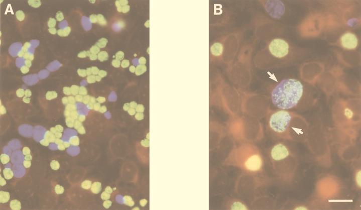
FIG. 1.
Dual infection of nonfusing (A) and fusion-competent (B) C. trachomatis strains cultured in HeLa cells and fixed for microscopy 30 h p.i. Within the nonfusing inclusions, serovar J(s) (green developmental forms) and serovar K(s) (blue developmental forms) remain segregated in individual vacuoles. Consistent with results of Ridderhof and Barnes (13), developmental forms of wild-type J (green) and K (blue) cells can be found within a single inclusion (arrows). The bar in panel B indicates 10 μm for both panels.

