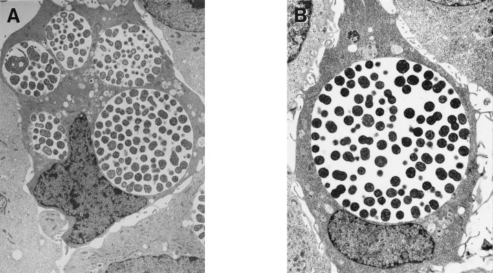
FIG. 2.
(A) Electron micrographs of HeLa cells infected with serovar J(s) EB at an MOI of 10 and fixed for electron microscopy 30 h p.i. Notice multiple vacuoles within single cells (magnification, ×6,200). (B) Parallel micrograph of HeLa cells infected with wild-type serovar J-infected HeLa cells (magnification, ×8,200).

