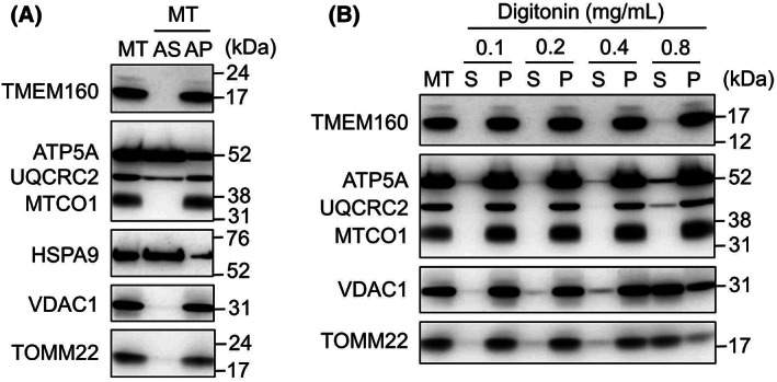
Fig. 2.

Confirmation of mitochondrial inner membrane localization of TMEM160. (A) Mitochondria were isolated from HepG2 cells as described in the “Materials and methods” and subjected to the alkali extraction assay. Immunoblot analysis was performed [AP, alkali‐resistant pellet fraction; AS, alkali‐soluble supernatant fraction; ATP5A, mitochondrial ATP synthase subunit alpha; HSPA9, mitochondrial Hsp70; MT, isolated mitochondria; MTCO1, mitochondrially encoded cytochrome c oxidase I; TOMM22, translocase of outer mitochondrial membrane 22; UQCRC2, ubiquinol‐cytochrome c reductase core protein 2; VDAC1, voltage‐dependent anion channel 1]. (B) the mitochondria isolated from HepG2 cells were treated with digitonin at the indicated concentration, followed by centrifugation to obtain the supernatant and pellet fractions [P, pellet fraction; S, supernatant fraction].
