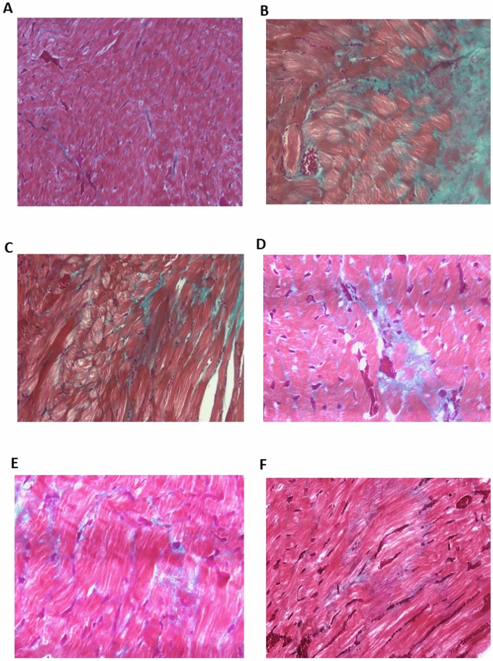
Figure 2.
Photomicrograph of heart tissue sections in different studied groups using Masson’s trichrome staining. (A) Control group exhibited a minimal amount of collagen fiber deposition around blood vessels (×200); (B) model group (dexamethasone-treated group) showed prominent and dense collagen fibrosis in the left ventricle (×400); fibrosis occurred in both interstitial and perivascular regions in an unorganized manner; (C) PRV 0.5 mg/kg; (D) PRV 1 mg/kg; (E) PRV 2 mg/kg with less fibrosis (×400). (F) Losartan treatment reduces cardiac fibrosis and collagen fibers are deposited around blood vessels (arrows) (×400).

