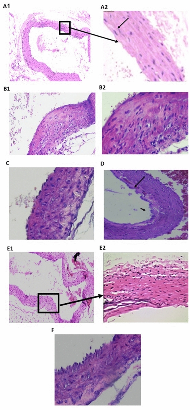
Figure 3.

Photomicrograph of aorta tissue sections in different studied groups. (H&E) (A) Control group: the tunica intima (arrowhead) faces the lumen. The tunica media is composed of elastic fibers and smooth muscle (thick arrow). The tunica adventitia is the outermost layer, which is composed of loose connective tissue (arrow) (A1,A2, respectively; ×100, ×200); (B1,B2) model group (dexamethasone- treated group) exhibited pathological changes, including lumen bulging and medial inflammatory cell infiltration (arrow), as well as an increase in the tunica media thickness overall (B1, ×200 and B2, ×400); (C) PRV 0.5 mg/kg there was a decrease in wall thickness associated with crystalloid material deposition (arrow); (D) PRV 1 mg/kg showed a decrease in wall thickness with organization of muscular fibers, with focal areas of disrupted endothelial cell lining fibrillation and focal subintimal hemorrhage (×200); (E) PRV 2 mg/kg with nearly normal endothelial lining and normal muscle structure (×200); (F) losartan-treated group with reduced intimal thickness.
